Standardize sample chicken bones used for x-ray testing
Testing an X-ray system is typically based on selecting small bones and cutting small pieces of these bones. These pieces are subsequently placed into a chicken fillet or a deboned leg. The product is subsequently scanned by the X-ray system, and its ability to detect the bone in question is assessed.
But a bone is not just a bone. The detectability of a bone depends on its calcification level, and the detection depends on the X-ray technology being used. Most important is the X-ray energy selection. At lower levels of X-ray energy and with longer wavelengths (below 30 kV), the contrast between bone and meat increases exponentially.
X-ray systems for poultry bone detection typically work around 60-80 kV. The high energy is needed because of the sensitivity limitation of standard X-ray imaging detectors. This is unfortunate, since the contrast between bone and meat reduces exponentially with increasing energy. For some years, research in the medical and dental sector has brought new detectors with very good imaging characteristics down to 10 kV. These are very interesting for chicken-bone detection, because the excellent contrast is matched with an excellent resolution.
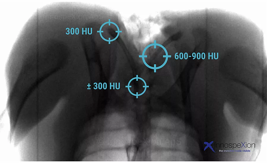
The quantitative distinction is based on differences in X-ray attenuation using the Hounsfield Unit (HU). This scale is based on water having the value of 0 HU, whereas air has the value -1,000 HU. A calcified bone from pork, for example, is typically in the range 600-1,000 HU (or beyond), whereas soft-bone (cartilage) is much lower (e.g. 100-250 HU), because of the lower abundance of calcium minerals.
For chicken, the bone HU value depends on the age of the chicken, its feeding, its breed and many other factors. For chicken in general food production, an average HU is about 400-450 HU for calcified bone, but varying from the level of cartilage (soft bone) (100-250 HU) up to 1,000 HU for the larger and most calcified bone.
Because the detection of a bone depends on the HU value of the bone, it makes no sense to consider only the dimension when evaluating the bone detectability. A comparison of different X-ray systems must therefore be made with the exact same bone pieces. This is difficult, since practical conditions may cause loss of the (very small) bone pieces used. The optimal solution therefore is to apply a test sample with artificial bone pieces (made of a polymer with hydroxyapatite) with a HU value (i.e. around 400-450) similar to that of the average chicken bone.
The above analysis unveils that the comparison of different X-ray systems should be based on standardized test samples. It is also clear that quality control verifications of X-ray system detection capability (as performed e.g. on an hourly regular) must be based on well-known, certified and calibrated test samples. It makes sense to use test samples with HU values in the range 400 to 450 HU, as these conform to the average range of calcified bones in general chicken production. NP
Looking for a reprint of this article?
From high-res PDFs to custom plaques, order your copy today!


